
Figure 3.
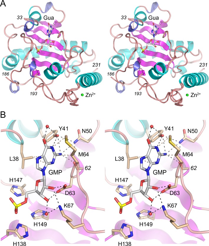
Structural basis for cap guanosine recognition by aprataxin. (A) Stereo view of the tertiary structure of the superimposed A and B aprataxin protomers, with β strands, α helices and 310 helices colored magenta, cyan and blue, respectively. Guanosine/GMP in the A/B active sites are shown as stick models. Zn2+ is depicted as a green sphere. (B) Detailed stereo view of GMP in the active site of the B protomer. Selected amino acids are shown as stick models with beige carbons.