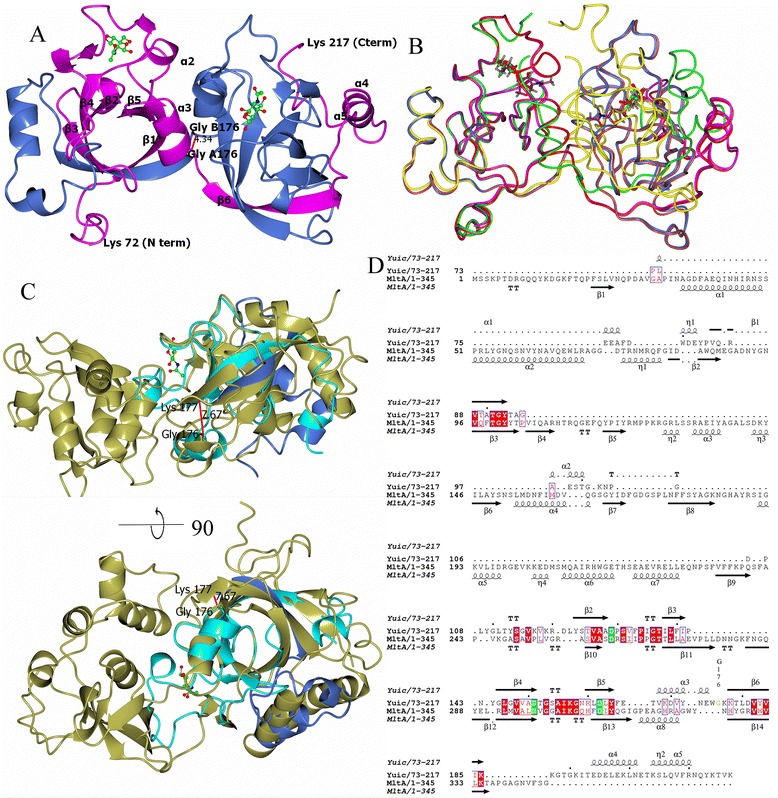
Fig. 1.

Structure of YuiC and comparison with MltA. a Dimer of YuiC with NAG bound. Chain A is in magenta and Chain B in blue with the positions of starts and ends of secondary structure elements labelled. NAG is ball and stick with carbon in green, oxygen in red and nitrogen in blue. The distance between the CA of G176 of each chain is shown in Å. b Structural Superposition of YuiC structure backbones. +NAG chains in magenta and blue with ligand in green, +Anhydro chains in red and pale crimson and ligand in dark purple. Apo chains in green and yellow. c Structural superposition of + NAG YuiC in cyan (A72-176) and blue (B177-217) (pseudo monomer) and MltA from E.coli (PDB 2ae0) [19] in gold. Lower picture is 90° rotation around horizontal of upper. The distance between A176 and B177 of YuiC is shown in Å. d Sequence alignment based on the structural superposition in C with secondary structure elements labelled, conserved aspartates shown in green and other conserved residues shown in red. G176, where the domain swap is centred, is coloured yellow and labelled. Structural superposition used SSM [36] in CCP4MG [37], structural alignment generated by UCSF chimera [38], structures drawn with CCP4MG [37] and alignment with ESPRIPT [39]
