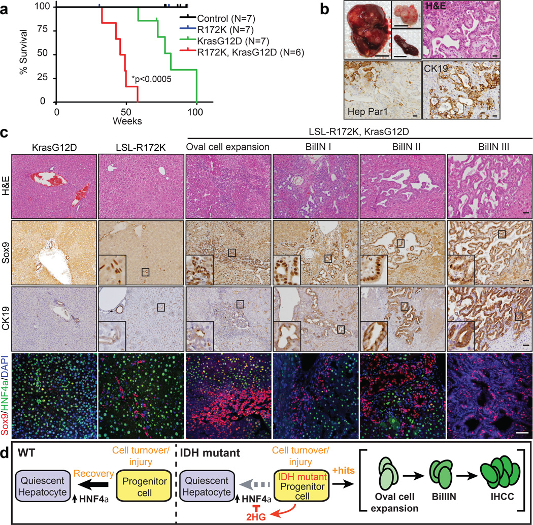
Figure 4. Mutant IDH cooperates with KrasG12D to drive liver progenitor cell expansion and multi-step IHCC pathogenesis.
a. Kaplan-Meier analysis showing time until signs of illness necessitated euthanasia. All animals euthanized had liver tumours.
b. Upper left: Representative Alb-Cre;LSL-R172K;KrasG12D tumor, and peritoneal and spleen metastases (insets). Upper right: H&E staining showing IHCC histology. Lower panels: The tumour is Hep Par1- and CK19+ while adjacent hepatocytes stain Hep Par1+ and CK19-.
c.The livers of Alb-Cre;LSL-R172K;KrasG12D animals exhibit oval cell expansion and increasing grades of BilIN, which stain Sox9+. CK19 levels increase with higher grade lesions. IF analysis reveals focal accumulation Sox9+ oval cells in Alb-Cre:LSL-R172K livers and pronounced oval cell expansion in Alb-Cre;LSL-R172K; KrasG12D livers.
d. Model for mutant IDH in cholangiocarcinoma pathogenesis. Scale bars, 1cm (b, upper left), 50µm (b-c).

