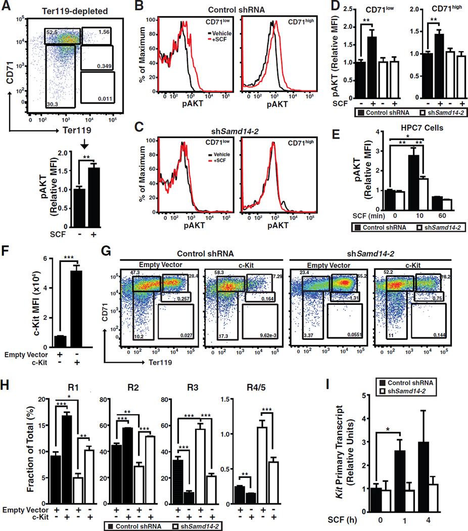
Figure 6. Samd14 Requirement for SCF/c-Kit Signaling.
(A) CD71 and Ter119 flow analysis of ex vivo expanded fetal liver cells following bead sorting for Ter119− cells. SCF-treated (10 ng/mL, 10 min) Ter119− cells analyzed for p-AKT MFI by flow cytometry. (B) p-AKT staining with control shRNA and CD71-low and CD-71-high fetal liver cells. (C) Phospho-flow with Samd14-knockdown CD71-low and CD-71-high fetal liver cells. (D) p-AKT MFI in control shRNA and shSamd14 fetal liver cells treated with 10 ng/mL SCF for 10 min (E) p-AKT MFI in control shRNA and shSamd14 HPC-7 cells treated with 50 ng/mL SCF for 0, 10 or 60 minutes. (F) c-Kit MFI in control and c-Kit overexpressed fetal liver cells. (G) Flow cytometry of enforced c-Kit expression, upon Samd14 knockdown, in fetal liver cells. (H) Percentage of cells in R1-R5 populations. (I) SCF treatment (50 ng/ml) of control or shSamd14-infected fetal liver cells 0, 1 and 4 h post-stimulation. Statistical significance: mean +/− SEM.; *p<0.05. **p<0.01, ***p<0.001.

