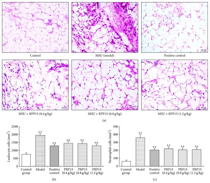
Figure 2.
Histomorphometric analysis of the effect of RPP15 on MSU-induced leukocyte and neutrophil infiltration. (a) Representative histological changes using HE stain of synovial tissue from rats knee joint 24 h after the injection of MSU crystals were shown (magnification, ×200). (b) Leukocyte and (c) neutrophil infiltration number were counted from HE stain of synovial tissue slides (cells per square millimeter). Data are means ± SD (n = 6). ++ P < 0.01 versus vehicle-control group (control); ∗∗ P < 0.01 versus vehicle with MSU crystal (model).

