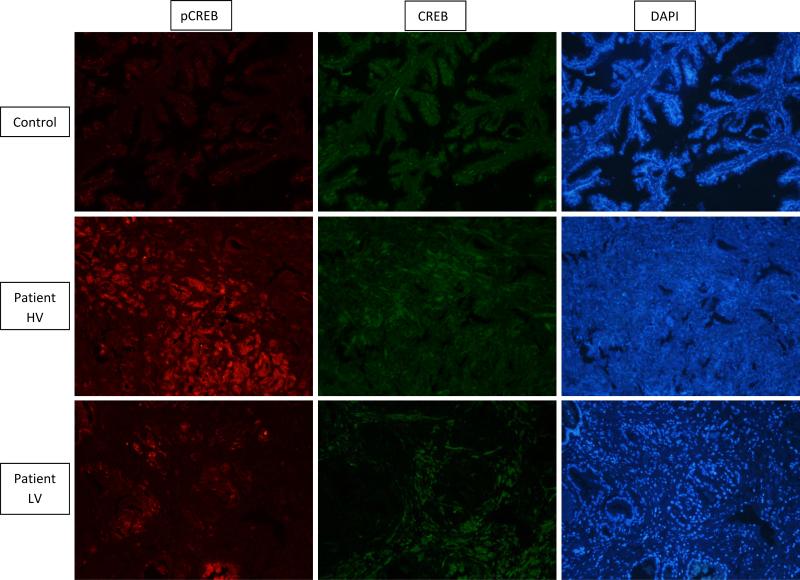
Figure 2.
Immunohistochemistry (IHC) for the CREB (green) and pCREB (red) proteins in prostate tissue (10x magnification). Primary antibodies of rabbit anti-CREB and mouse anti-pCREB (dilution 1:100) and secondary antibodies goat anti-rabbit 488 and goat anti-mouse 555 where used to achieve this image. Higher levels of pCREB (red) are observed in PCa tissue when compared to a control; there does not appear to be a difference in unphoshorylated CREB (green). Patient (HV) – High number of PDE variants; Patient (LV) – Low number of PDE variants.

