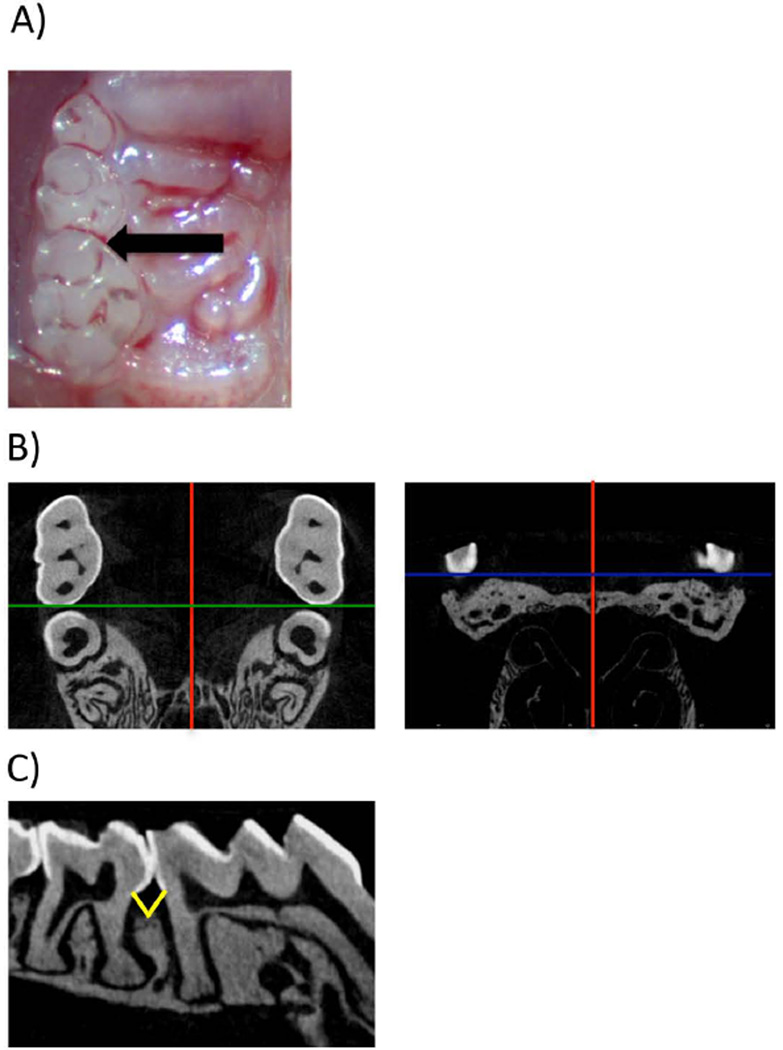
Figure 1.
Injections and Micro-CT image/sample orientation. A) Clinical image with the location of LPS injection. B) Micro-CT data were imported in DICOM format into Dolphin® software and the image volume was oriented in the orthogonal planes such that red line denotes (sagittal plane), green line (coronal), blue line (transverse plane): B1) the axial slices are parallel to the occlusal plane. The intermaxillary suture is parallel to the sagittal plane. C) The distance from the CEJ to the alveolar crest was measured at the sagittal plane intersecting the interproximal molars. Yellow lines depict the measurement that was taken for distal of 1st molar and mesial of 2nd molar.

