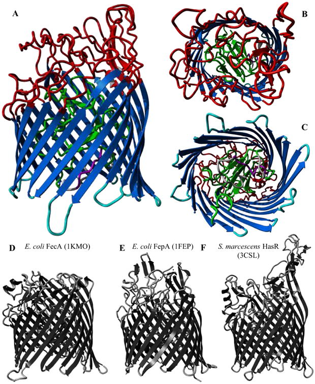
Figure 3.

A-C: Predicted structure of HemR from Finnish OM isolate F433-3 (C-score = 0.74). Structural domains are colored as follows: gray – signal peptide; purple – putative TonB box; green – N-terminal plug domain; dark blue – transmembrane β-strands; red – extracellular loops; light blue – intracellular loops. A. Side view. B. Extracellular side. C. Periplasmic side. D-F: Experimentally derived structures for three iron acquisition receptors in other gram negative species, with PDB IDs in parentheses. D. E. coli ferric citrate uptake transporter FecA. E. E. coli ferric enterobactin receptor FepA. F. S. marcescens hemophore receptor HasR.
