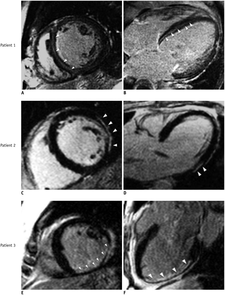
Fig. 1. Different delayed enhancement patterns in non-ischemic cardiomyopathy.
Patient 1. 48-year-old female with history of dilated cardiomyopathy. Delayed enhancement images demonstrate intramural contrast enhancement in septum (midwall striae, arrowheads). A. Mid-ventricular short-axis view. B. 4-chamber-view. Patient 2. 50-year-old male with remote history of biopsy-proven viral myocarditis presented with left ventricular dysfunction. Delayed enhancement images demonstrate epicardial hyperenhancement localized at basal inferolateral wall (arrowheads). C. Basal-short axis view. D. 3-chamber-view. Patient 3. 68-year-old male presented with progressive left ventricular dysfunction found to have insignificant coronary stenosis (25% middle left anterior descending artery lesion) on invasive coronary angiography. Delayed enhancement images demonstrate subendocardial hyperenhancement with 75% transmurality (arrowheads) involving inferior and inferolateral walls from middle ventricular level extending through apex consistent with ischemic injury. E. Mid-ventricular short axis view. F. 3-chamber-view.

