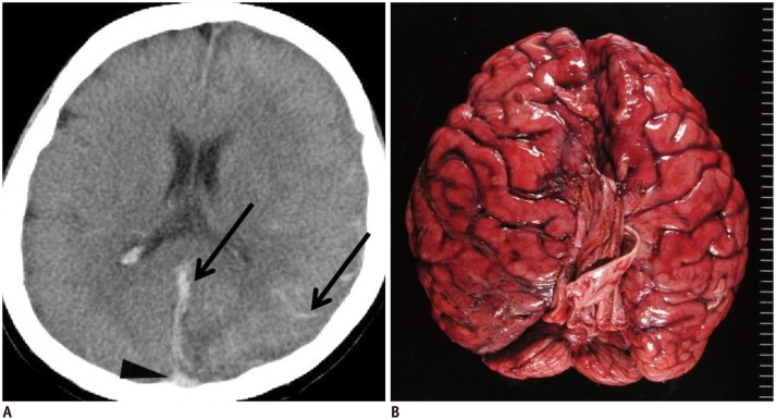
Fig. 1. Hypostasis and hemorrhagic lesion in brain in 35-year-old deceased woman (case 1).
A. CT scan obtained 8 hours and 35 minutes after death shows obscure hypostasis in dorsal superior sagittal sinus (arrowhead) in case 1. Linear and curving high density lesions are present along falx cerebri and cerebral sulcus (arrows). B. Subsequent autopsy reveals diffuse subarachnoid hemorrhage. CT = computed tomography

