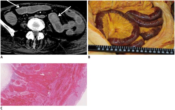
Fig. 11. Hyperdense wall of GI tract in 74-year-old deceased woman (case 13).
A. CT scan obtained 2 hours and 34 minutes after death shows hyperdense walls throughout GI tract (arrows). B, C. Autopsy revealed that hyperdense GI wall was intramural hemorrhage (B, macroscopic image; C, microscopic low-power view image with hematoxylin and eosin stain). CT = computed tomography, GI = gastrointestinal

