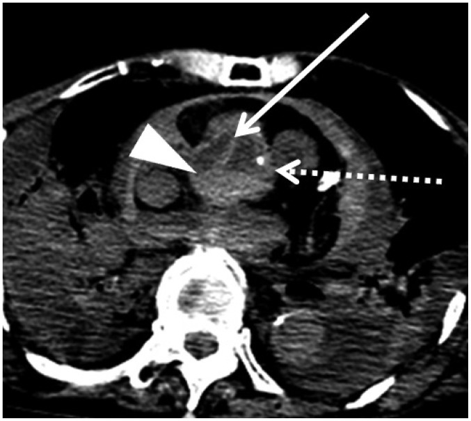
Fig. 4. Dissection and hypostasis in ascending aorta in 76-year-old deceased woman (case 6).
CT scan obtained 14 hours and 30 minutes after death shows both flap (arrow) separating two lumens and hypostasis (arrowhead) in ascending aorta. Increased density and thickening of aortic wall, which are normal postmortem changes, are also observed (dotted arrow). CT = computed tomography

