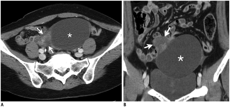
Fig. 7. Contrast-enhanced computed tomography (CT) scan in 38-year-old woman with complaint of acute pelvic pain as false-positive case.
Transverse CT scan (A) and coronal reformation (B) show well-defined cystic mass (*) with eccentric soft tissue lesion (arrows). Both readers provided score of 4 as level of suspicion for adnexal torsion using 5-point scale. Adnexal mass was paratubal cyst without torsion in right ovary.

