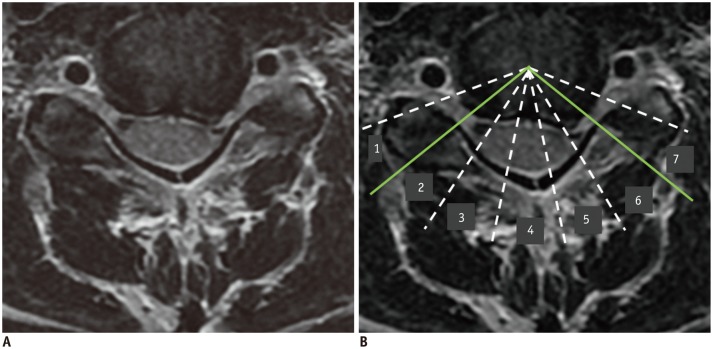
Fig. 3. Epicenter and number of involved segments (NIS) of herniated disc on MRI.
45-year-old female with left upper extremity tingling sensation. A. T2-weighted axial imaging shows disc protrusion at C5-6 level. B. Eight virtual lines delimit seven segments, and epicenter (6) and NIS (5, 6, and 7; i.e., three segments) of herniated disc material are determined.

