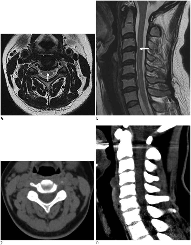
Fig. 4. Comparison of cervical MRI and MDCT in 43-year-old female with posterior neck pain.
Central disc protrusion is evident at C3-4 level on MRI (A, B; arrows). However, all readers interpreted disc as normal on MDCT (C, D). MDCT = multidetector-row computed tomography, MRI = magnetic resonance imaging

