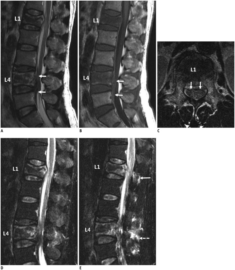
Fig. 4. Case of disagreement about posterior ligamentous complex (PLC) integrity by readers.
51-year-old man demonstrated traumatic lesions at L1 and L4 bodies on T2- (A) and T1-weighted sagittal images (B) with epidural hemorrhage at L3-5 level (arrows, A, B). L1 lesion revealed bulging of posterior cortex on T2-weighted axial scan (C, arrows), which was agreed to be burst injury by all readers during first review and by five during second review. However, no consensus was reached about PLC integrity on T2-weighted short time inversion recovery sagittal images (D, E, arrow), which produced variety of scores for PLC intergtity ("intact" by two readers; "indeterminate" by four in first and "intact" by three; "indeterminate" by two; and "injured" by one during second review). In contrast, PLC integrity (E, dashed arrow) of L4 body lesion was not evaluated using thoracolumbar injury classification system and was originally classified as most severe injury level.

