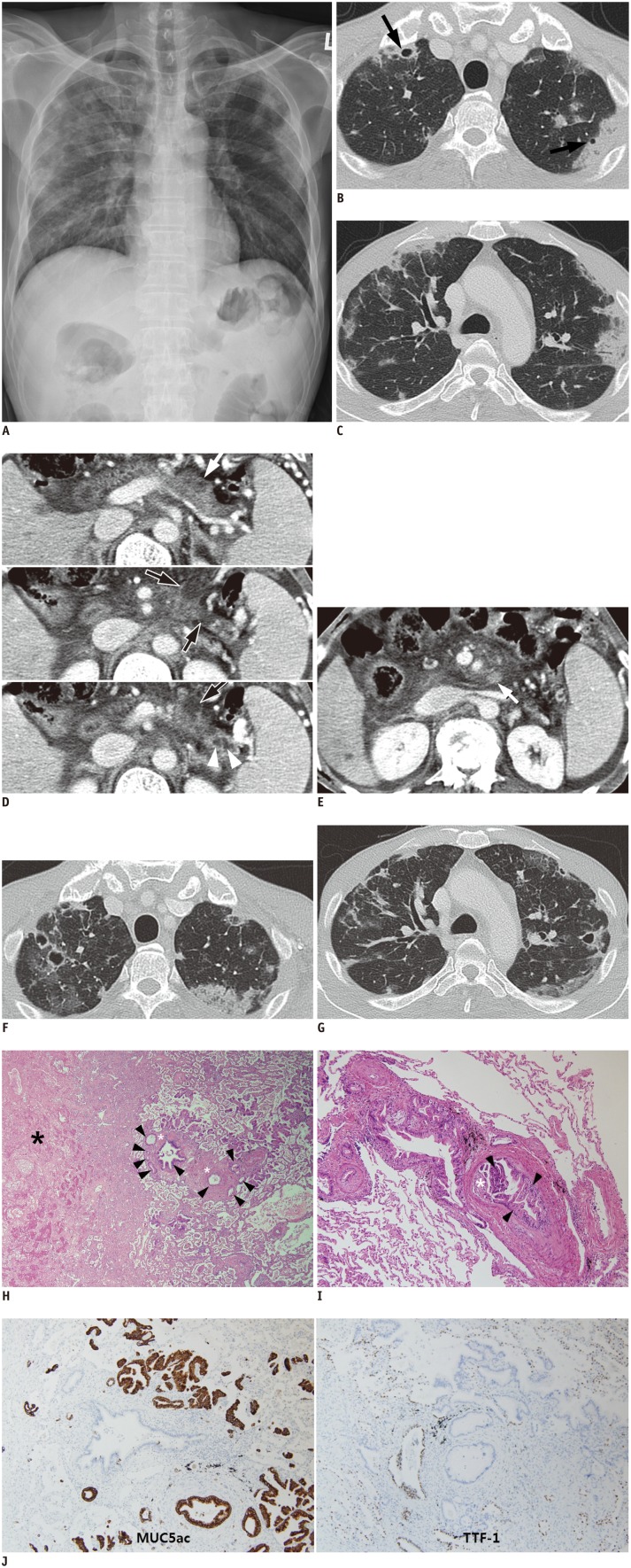
Fig. 1. 52-year-old man with cavitary infarction caused by pulmonary tumor thrombotic microangiopathy originated from pancreatic intraductal papillary mucinous neoplasm.
A. Chest radiograph demonstrates bilateral multifocal consolidations in peripheral portions of both upper lungs. Axial computed tomography images (lung window setting; window width, 1500 Hounsfield unit [HU]; window level, -700 HU) show ill-defined wedge-shaped consolidations and ground glass opacities in peripheral portion of both upper lobes (B, C). Cavitation was present in some consolidations (B, arrows). D. Contiguous contrast-enhanced axial CT images show multiple cystic lesions (black arrows with white border) with minimal disproportional pancreatic duct dilatation (branch ducts, white arrowheads) in body and tail of pancreas. Ill-defined soft tissue lesion is present in one cyst (white arrow), which is presumed to be mural nodule of malignant intraductal papillary mucinous neoplasm. E. Lymphadenopathy in peripancreatic space (arrow) and ascites are also seen. F, G. Follow-up axial CT images (lung window setting: window width, 1500 Hounsfield unit [HU]; window level, -700 HU) 3 weeks after initial CT scan demonstrate that wedge-shape consolidations are extended or newly developed. Additionally, cavities in consolidations are newly developed or changed, predominantly in peripheral portion of lungs. H. Photomicrograph of histopathological specimen shows tumor emboli (black arrowheads) and intimal hyperplasia (white asterisks) in vasculature. These tumor emboli and intimal hyperplasia may obstruct vascular lumen and cause irregular vascular shape. Distal lung parenchyma of obstructed vascular lumen is necrotic (black asterisk) (hematoxylin-eosin, original magnification, × 40). I. Photomicrograph of histopathological specimen shows tumor cells, columnar mucin-producing glands (black arrowheads) in pulmonary vasculature, and marked fibrocellular intimal hyperplasia (white asterisk) (hematoxylin-eosin, original magnification, × 100). There is little fibrin thrombosis in vascular lumen. J. Immunohistochemical stain for MUC5ac (× 100), to which metastatic adenocarcinomas demonstrate positive reaction (brown color) but bronchial epithelium and alveolar pneumocytes are negative. In contrast to MUC5ac, basal cells of bronchial epithelium and alveolar pneumocytes demonstrate positive reaction (brown color) to immunohistochemical stain for TTF-1 (× 100), but metastatic adenocarcinomas are negative.

