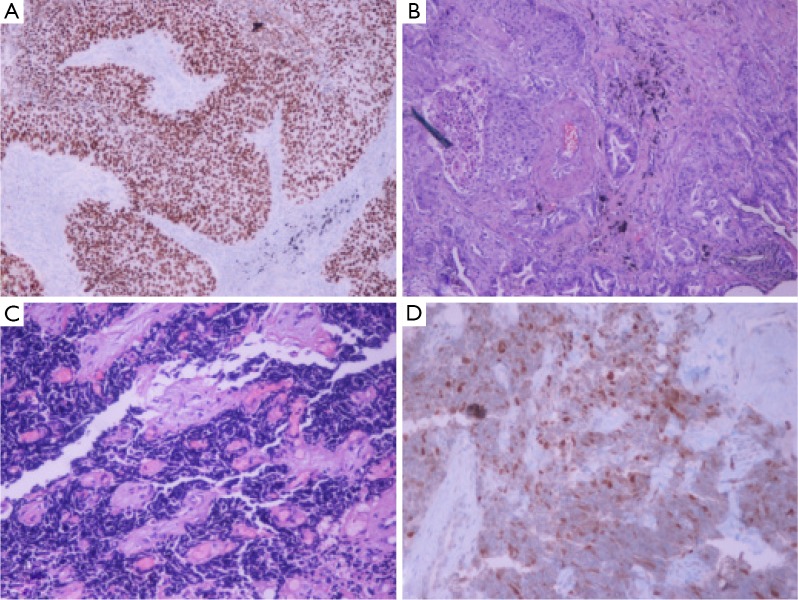
Figure 2.
Histology figures. (A) Squamous cell carcinoma, P63 immunostaining, original magnification ×20; (B) adenosquamous carcinoma, H-E stain, Presence of neoplastic glands and squamous cell nests, original magnification ×20; (C) small cell lung cancer (SCLC), H-E stain, fibrous tissue infiltrated by SCLC, original magnification ×20; (D) SCLC, thyroid transcription factor-1 (TTF-1) immunostaining, original magnification ×40.

