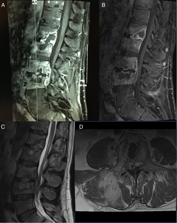
Figure 2.
Sagittal views of the lumbar spine demonstrate a heterogeneous mass with diffuse signal alterations in the L1 vertebra extending into L2 and in L3 extending into L4 with enhancement of the vertebral bodies (A). Repeat images again reveal destruction of L1–L4 with small epidural fluid collection communicating between superior aspect of L3 through inferior aspect of L4 and extending into right L3–L4 neural foramen (B–D).

