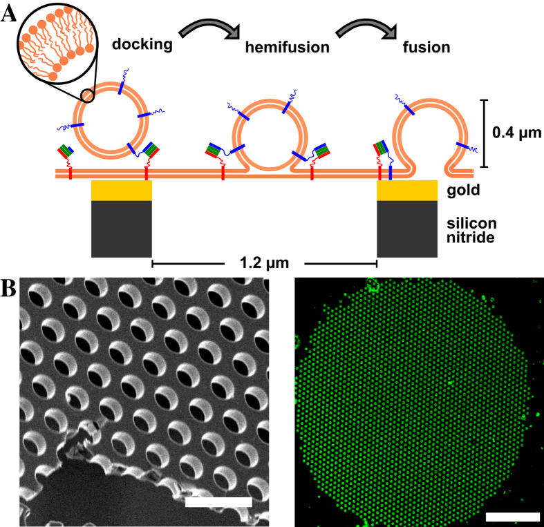
Figure 1.
(A) Schematic drawing of the model system for SNARE mediated membrane fusion based on pore-spanning membranes. SNARE proteins are shown in red (syntaxin 1A), green (SNAP25) and blue (synaptobrevin 2). Membranes are depicted as thick orange lines. (B) Scanning electron micrograph of a porous silicon nitride substrate. Scale bar: 3 μm. (C) Pore-spanning membrane patch obtained from spreading a giant unilamellar vesicle composed of DOPC/POPE/POPS/cholesterol (5:2:1:2) and doped with 1 mol % Oregon Green DHPE on a gold/6-mercapto-1-hexanol-functionalized substrate. Scale bar: 20 μm.

