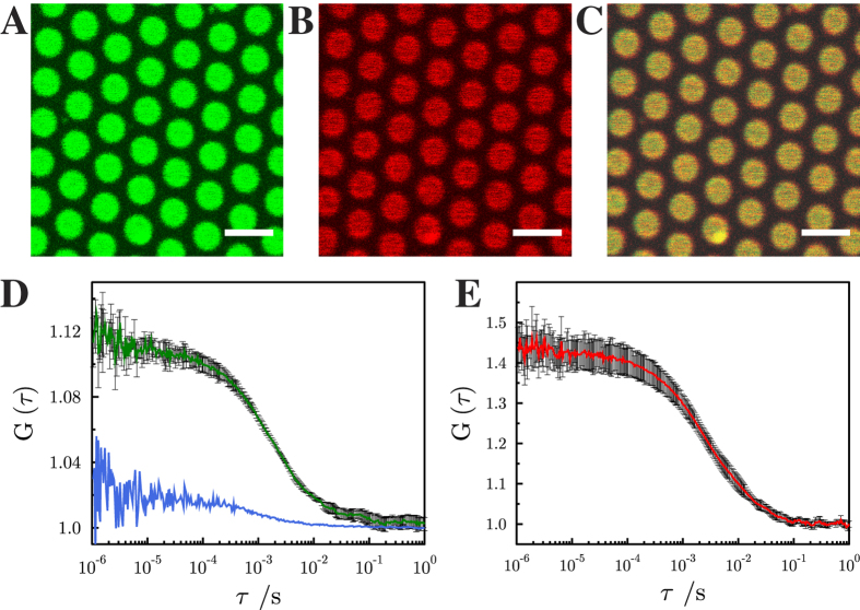
Figure 2. Fluorescence micrographs of pore-spanning membranes composed of DOPC/POPE/POPS/cholesterol (5:2:1:2) on a 6-mercapto-1-hexanol functionalized gold covered porous silicon nitride surface.
The images show fluorescence signals of (A) Atto488 DPPE, (0.01 mol%), (B) Atto647N-syntaxin 1 transmembrane domain (0.0055 mol%) and (C) an overlay of (A) and (B). Scale bars: 3 μm. Autocorrelation curves of the performed FCS measurements with excitation wavelengths of 488 nm (Atto488 DPPE, green, D) and 633 nm (Atto647N-syntaxin 1-TMD, red, E). The blue FCS curve in D was obtained on a gold covered pore rim showing that the fluorescence of Atto488 DPPE is significantly quenched and hence, the diffusion constant cannot be determined. Fitting eq. (4) to the autocorrelation curves provide diffusion coefficients of 7.7 ± 0.4 μm2/s (SD) for Atto488 DPPE and 3.4 ± 0.2 μm2/s (SD) for Atto647N-syntaxin 1-TMD. For Atto488-DPPE L was 200 nm, and for Atto647N-syntaxin 1A L was 250 nm. For each diffusion constant, at least 20 pore-spanning membranes from three independent reconstitutions were analyzed.

