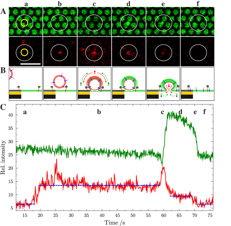
Figure 3.
(A) Time lapse series of fluorescence micrographs showing a single fusion event of a large unilamellar vesicle containing full length synaptobrevin 2 with a pore-spanning membrane with reconstituted ΔN-acceptor complex. (top) Oregon Green DHPE fluorescence of the pore-spanning membranes; (bottom) Texas Red DHPE fluorescence of the vesicle. The yellow circles in (a) shows the region of interest (ROI) that was used for reading out the time-resolved changes in the relative fluorescence intensities depicted in (C); the white circles in the images highlight the region, where the vesicle docks and fuses and only serves as a guide to the eye. Scale bar: 5 μm. (B) Schematic drawing of the postulated fusion states at the time of image recording. Membranes are colored according to their incorporated fluorescent dye. (C) Time resolved changes in the relative fluorescence intensity of Oregon Green DHPE (green) and Texas Red DHPE (red) during the shown fusion event. Dashed blue lines serve as a guide to the eye highlighting the distinct levels of intensity. Letters (a–f) correspond to the fluorescence images in (A). For further details see text and Supplementary Fig. 7.

