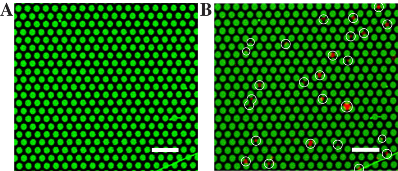
Figure 7.

Fluorescence micrographs of a pore-spanning membrane patch containing cholesterol-PEG12-(EIAALEK)4 (A) before and (B) after the addition of large unilamellar vesicles containing cholesterol-PEG12-(KIAALKE)4. All 26 Texas Red DHPE doped vesicles, marked with white circles, have docked onto the pore-spanning membranes at the end of the time series. Scale bars: 5 μm.
