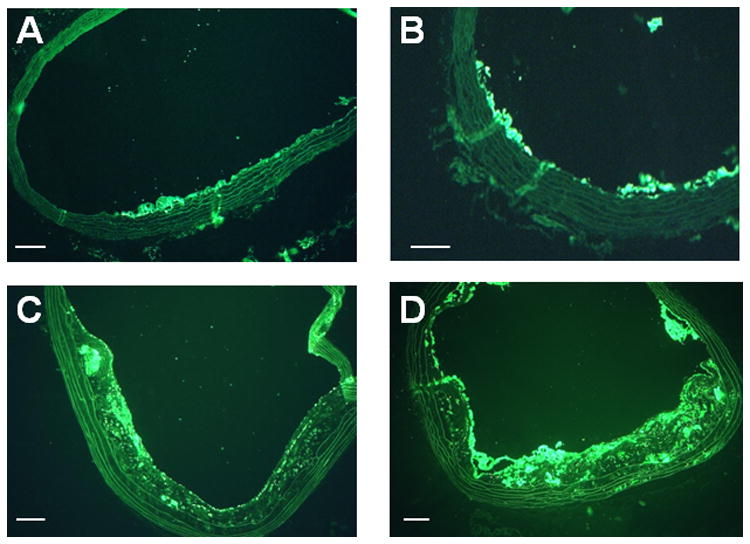
Figure 1.

Histology from DKO mice. Mac-2 immunohistochemistry illustrates increasing degree of plaque size and plaque macrophage content with increasing age of DKO mice from (A) 10 weeks, (B) 20 weeks, (C) 30 weeks, and (D) 40 weeks of age. Masson's trichrome stain shows increasing plaque size from DKO mice at (E) 20 weeks, (F) 30 weeks, and (G) 40 weeks of age. (H) Example of immunostaining of β3-integrin from a 40 week DKO mice illustrating the presence of platelets on the plaque luminal surface (arrows). Endothelial staining presumably from αv β3 is seen as well. Scale bars = 100 μm.
