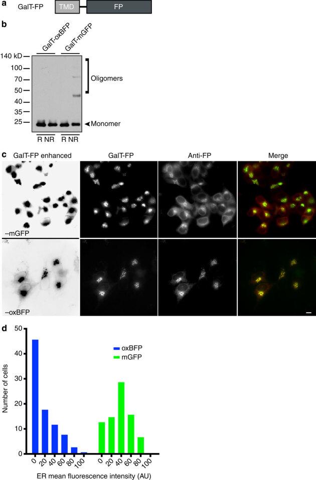
Figure 4. Golgi complex membrane-localized mGFP forms inappropriate disulphide bonds and is inefficiently trafficked to the GC.

(a) Schematic of GC-localized FP (GalT–FP) containing GalT transmembrane domain upstream of FP. (b) Immunoblot revealed the tendency of GalT–mGFP to form oligomers under NR conditions. Optimized, cysteine-less GalT–oxBFP does not form inappropriate disulphide bonds. (c) Representative images of HeLa cells transiently transfected with GalT–mGFP or –oxBFP. Immunofluorescence with anti-GFP revealed a significant fluorescently undetectable pool of GalT–mGFP in the ER. ER labelling by the FP is digitally enhanced with Levels tool in Photoshop in far left panels. Note that weak ER is apparent in all of the oxBFP expressing cells, but rarely observed in mGFP expressing cells. Scale bar, 10 μm. (d) Distribution of the ER fluorescence intensity values (mean fluorescence intensities of regions of anti-GFP staining proximal to the GC). n≥80 cells collected from 11 to 13 fields per construct.
