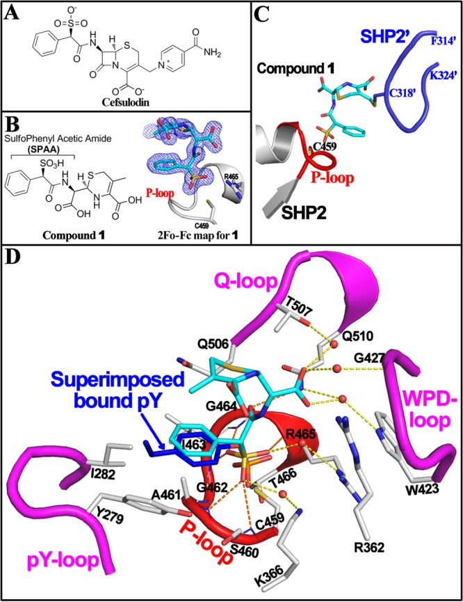
Figure 1.

Crystal structure of SHP2 with cefsulodin derived compound 1 identifies SPAA as a novel pTyr mimetic. (A) Structure of cefsulodin. (B) Structure and electron density of compound 1 bound at SHP2 active site. (C) Under crystallization conditions, the bound compound 1 (cyan) is covalently attached to C318 in a flexible loop (blue) from a nearby symmetric SHP2. (D) Interaction details of compound 1 (cyan) with SHP2. Residues within 5 Å distance of compound 1 were shown in stick (gray). H-bonds or water-bridged H-bonds between compound 1 and SHP2 were indicated by dash line. Four loops forming SHP2 active site pocket were highlighted in red or magenta. A PTP1B·pTyr-peptide structure (PDB#: 1EE0) was superimposed onto the SHP2·1 structure, and the bound pTyr (blue) was found to overlap well with SPAA.
