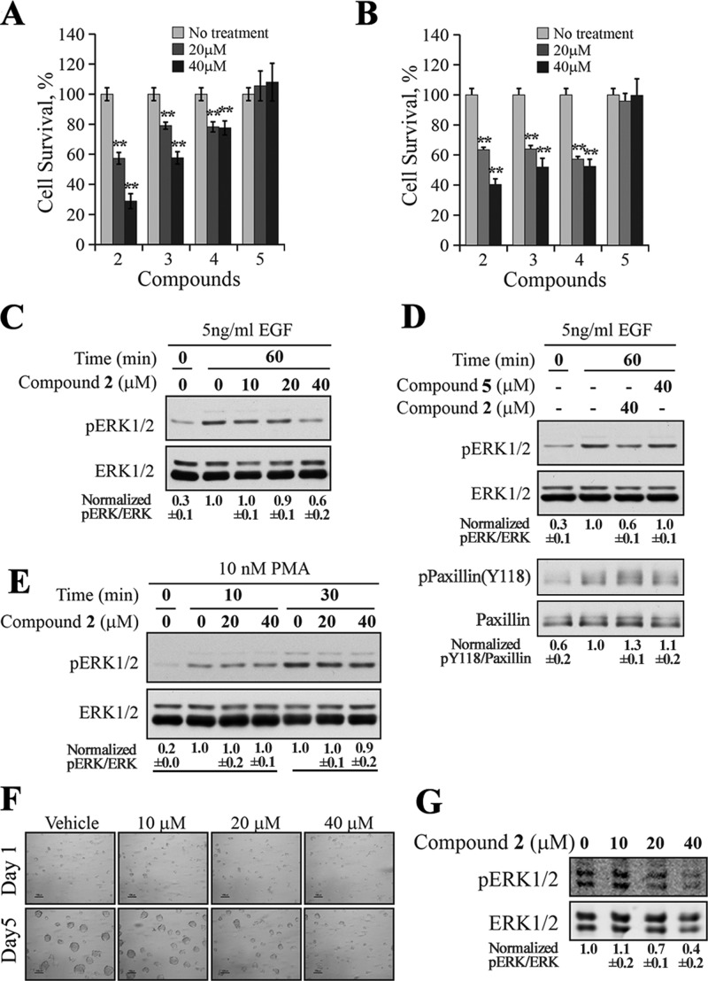
Figure 5.
Cellular activity of SPAA-based SHP2 inhibitors. MTT assay for compounds 2 to 5 in H1975 lung cancer cell line (A) or in MDA-MB-231 breast cancer cell line (B). Compounds 2–4 significantly (**p < 0.01) reduced cell proliferation in a dose-dependent manner in both cell lines. (C) Compound 2 decreased the EGF-induced ERK1/2 activation dose dependently in H1975 cell. (D) Compound 2 blocked EGF-induced ERK1/2 activation and SHP2-mediated dephosphorylation of paxillin, while the negative control compound 5 failed to exert any effect on ERK1/2 and paxillin phosphorylation. (E) Compound 2 had no effect on PMA-induced ERK1/2 activation. (F) Compound 2 inhibited the growth of the ErbB2 positive SKBR3 cells in 3D Matrigel. (G) Compound 2 inhibited ERK1/2 activation in SKBR3 cells cultured in a 3D Matrigel environment. The results shown in this figure are representatives from at least two independent experiments, and the numbers below the gels are presented as mean ± SD. The quantification and normalization were performed as follows: band intensity was quantified using the ImageJ program, and the ratios of pERK1/2/total ERK1/2 or pPaxillin(Y118)/total Paxillin were calculated and normalized to the reference.

