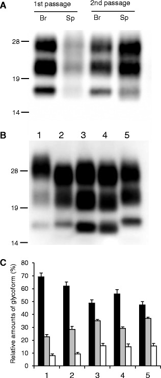
Figure 3.

Western blot analysis of proteinase-K resistant PrP Sc analyzed using monoclonal antibody T2. A PrPSc in the brain (Br) and spleen (Sp) of wild-type mice inoculated with sheep-passaged L-BSE at the first and second passage. All samples were digested with 50 μg/mL of proteinase-K at 37 °C for 1 h. Lanes from left to right were loaded with 0.625, 5, 0.0125, and 0.36 mg tissue equivalent, respectively. The molecular markers are shown on the left (kDa). B PrPSc in the brain of C-BSE- and L-BSE-affected cattle and mice. Lane 1: C-BSE affected cattle, Lane 2: C-BSE affected ICR mouse, Lane 3: L-BSE affected cattle, Lane 4: L-BSE affected sheep, and Lane 5: sheep-passaged L-BSE affected ICR mouse at second passage. Lanes 1, 3, and 4, and Lanes 2 and 5 were loaded with 1.25 and 0.125 mg tissue equivalent, respectively. C Quantification of the relative amounts of the di-, mono-, and unglycosylated forms of PrPSc from the brain. The column numbers are as listed in (B). Bar diagram indicates the diglycosylated form (black), monoglycosylated form (gray), and unglycosylated form (white). Data are expressed as mean ± standard deviation of triplicate experiments.
