

Abstract
[Purpose] This study aimed to evaluate the anti-inflammatory and analgesic effects of intraoral application of low-level laser therapy (660 nm) to control pain, swelling and interincisal opening following the extraction of mandibular third molars. [Subjects and Methods] Ten patients underwent removal of lower third molars using the same surgical protocol and pharmacological approach. In the postoperative period, all patients received four consecutive daily sessions of low-level laser therapy, beginning 24 hours after the surgery. Intraoral applications using the diode laser with 660 nm wavelength in the continuous scan mode were performed covering the entire surgical area, which was divided into four quadrants, each of 1 cm2 area at a distance of 1 cm. The energy applied at each point was 5 J/cm2 during 8 seconds. [Results] The swelling and interincisal opening returned to normal 24 hours after the first low-level laser therapy application (Friedman test). Moreover, the pain intensity was reduced on the third postoperative day, according to the Friedman test. [Conclusion] Low-level laser therapy (660 nm), at the dosimetry used in this study, was effective in reducing postoperative pain and swelling following oral surgery.
Key words: Low-level laser therapy, Rehabilitation, Inflammation
INTRODUCTION
Removal of third molars is a common procedure in oral surgery and it is associated with postoperative complications such as pain, swelling and trismus. Therefore, several medications such as corticosteroids and non-steroidal anti-inflammatory drugs are often recommended despite their common side effects such as bleeding, gastrointestinal irritation and allergic reactions, which can evoke discomfort and health risks in patients1).
In terms of strategies used to minimize tissue damage in oral surgeries, the low-level laser therapy (LLLT) seems to offer many benefits in the stimulation of the healing process and control of the inflammatory process by reducing the swelling and pain, with no reports of adverse effects2).
According to Medeiros et al.3), the effects of LLLT are more evident in the early stages of wound healing. The use of polarized light helps in resolution of inflammation and in increasing the collagen deposition as well as the number of myofibroblasts. Moreover, it also fastens re-epithelialization during the experimental period. However, previous studies have reported that in teenagers, LLLT reduces pain intensity without modifying the healing process4). On the other hand, LLLT showed better results in controlling swelling and trismus when compared to ozone therapy5). However, Lopez-Ramirez et al.2) found that LLLT showed no positive effects in reducing pain, swelling and trismus after the removal of third molars.
These differences in the effects of LLLT may be explained by the variability in the protocols and surgical procedures performed1, 2, 6).
The LLLT protocol should be determined considering the type of light used as well as the tissue layers that need to be irradiated. In this context, the red spectra in the visible to near-infrared spectra (620–830 nm) have the potential to stimulate photoreceptors in the plasma membranes of cells. The lower wavelengths are mainly absorbed by superficial tissues with red pigmentation. The higher wavelengths are absorbed by the deeper tissues and are less active in the surface tissues7). Therefore, this study aimed to evaluate the efficacy of intraoral application of LLLT (660 nm) in controlling pain intensity, swelling and interincisal opening in oral surgery.
SUBJECTS AND METHODS
This study included 10 patients (7 women, 3 men, mean age 23 ± 6 years) and it was initiated after obtaining the approval of the Ethics in Research Committee (Protocol number: 512.476). All patients have signed a written informed consent prior to any procedure. The exclusion criteria were as follows: presence of local infections, various systemic diseases, smoking habit, use of oral contraceptives, pregnancy and lactation.
All patients underwent the same surgical protocol standardized by the same surgeon (R.L.N.). The same operator applied LLLT in all patients (H.S.C.F.). However, the values recorded for pain, swelling and trismus were assessed by a different surgeon in order to conduct a blind experiment. Before the laser application, the mean power of the equipment was measured and calibrated using the laser check power meter instrument.
Intraoral LLLT was applied using continuous scan mode at four different points, covering the entire suppurative surgical area, which was divided into four quadrants, each of 1 cm2 area at a distance of 1 cm.
Preoperatively, all patients used a mouthwash with chlorhexidine (0.12%) and received a topical application of povidone-iodine (1%). Postoperatively, all patients received amoxicillin 500 mg, every 8 hours, for 7 days, ibuprofen 600 mg, every 8 hours, for 3 days, and paracetamol 500 mg, every 6 hours as rescue medication for pain during the first 72 hours. In cases with penicillin allergy, clindamycin was selected as the antibiotic at a dose of 300 mg, every 8 hours, for 7 days.
In this study, the laser model Endophoton LLT 0107 (KLD Biosystems Electronic Equipment Ltd., Amparo, SP, Brazil) with a pen station beam area of 0.035 cm2, AlGalnP visible diode with continuous wave (35 mW and wavelength of 660 nm) was used. The energy applied at each point was equal to 5 J/cm2/dose, continuous rate for 8 seconds, as specified by the manufacturer.
The formula below was used to calculate the energy density (D), where the area (A: cm2) corresponds to the broadcast area of the pen tip, power (P: W) corresponds to the maximum and constant power (20 mW or W 0.020) and the time (T) is measured in seconds.
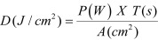 |
 |
After applying the values supplied by the manufacturer in the formula, the energy density applied at each point was calculated to be 4.57 J/cm2 multiplied by 4 quadrants of 1 cm2, resulting in a total of 18.28 J/cm2 in each session. Considering previous reports of several authors, a dose of 3 J/cm2 up to 6 J/cm2, 8, 9) has a regenerative action.
Four consecutive daily sessions, with the first session occurring 24 hours after surgery, were applied. This protocol was chosen based on the fact that the process of wound repair is composed of 3 phases: inflammatory, proliferative, and remodeling phases involving the formation of collagen10), and LLLT is more evident in the initial stages of healing2). However, in this protocol, the acute phase of inflammatory process is avoided.
The outcome variables assessed were pain, interincisal opening, facial swelling, and presence of late infection. The experimental times considered in this protocol were as follows: t0 (initial time), t1 (24 h), t2 (48 h), t3 (72 h), t4 (96 h), t5 (120 h), t6 (144 h), and t7 (7 days after the surgery).
The interincisal opening was assessed by measuring with a caliper of maximum opening between the right upper and lower central incisors before surgery and in all subsequent sessions11).
To evaluate swelling, a measurement from one tragus to the opposite one was performed, according to method described by Schultz-Mosgauet et al12).
All patients were instructed to register the daily intensity of pain, beginning 24 hours after surgery, for seven postoperative days, using a visual analogue scale (VAS) of 100 mm, in which the score for lack of pain was 0 (zero), and 100 (one hundred) was the score used for the worst imaginary pain.
All data were analyzed using GraphPad 5.0 (GraphPad Software, La Jolla, California, USA). The level of significance was set at p < 0.05 with a confidence interval of 95%.
RESULTS
This study included 10 patients (mean age: 23 ± 6.03 years) with asymptomatic third molars (no pain observed on the day prior to the surgery, as assessed by VAS). The average operative time was 29.45 ± 12.67 minutes. All data on the interincisal opening, swelling and pain intensity are shown in Table 1.
Table 1. All data on the interincisal opening, swelling, and pain intensity.
| Post-operatory evaluation | T0 | T1* | T2 | T3 | T4 | T5 | |
|---|---|---|---|---|---|---|---|
| Interincisal openning (mm) | 25th. quartile | 45.0 | 32.2 | 36.7 | 33.5 | 38.0 | 43.5 |
| Median | 47.5 | 37.0 | 43.5 | 43.0 | 44.0 | 48.0 | |
| 75th. Quartile | 50.0 | 45.2 | 46.5 | 48.0 | 48.2 | 51.2 | |
| T0 | T1** | T2 | T3 | T4 | T5 | ||
| Swelling assessment (mm) | 25th. quartile | 271.5 | 271.5 | 273.3 | 273.0 | 271.0 | 273.5 |
| Median | 280.0 | 283.5 | 283.5 | 280.5 | 279.5 | 283.5 | |
| 75th. Quartile | 289.0 | 296.3 | 289.8 | 288.3 | 289.8 | 290.0 | |
| T0 | T1 | T2 | T3*** | T4*** | T5*** | ||
| Pain intensity (100 mm) | 25th. quartile | 24.5 | 25.2 | 23.0 | 13.7 | 6.0 | |
| Median | 49.0 | 40.0 | 28.5 | 23.0 | 16.5 | ||
| 75th. Quartile | 70.2 | 54.0 | 45.0 | 29.7 | 25.2 | ||
* Statistically different from T0, Friedman Test, p<0.05. ** Statistically different from T0, Friedman Test, p<0.05. *** Statistically different from T0, Friedman Test, p<0.05.
With regard to the interincisal opening, a statistical decrease was observed 24 hours after the surgery (Friedman test, p = 0.0001). Similarly, an increase in swelling was observed when comparing the initial condition and the first postoperative day (Friedman test, p = 0.01). Therefore, it may be noted that both the swelling and interincisal opening returned to normal condition 24 hours after the first LLLT application.
Concerning the pain intensity (assessed by VAS), no differences were observed between the first and second postoperative days (median: 49 versus median: 40). However, a reduction in pain intensity was observed immediately after the third postoperative day, reinforcing the possible effect of LLLT on pain (Friedman test, p = 0.001). Additionally, on the seventh day, the patients were asymptomatic. Even though the patients with a greater surgical duration had a slight increase in pain intensity on the first day, it was not statistically significant (Spearman’s correlation, p > 0.05). Additionally, none of the analyzed cases had any late complications.
DISCUSSION
In this case series, we have focused on the local postoperative clinical factors after removal of third molars, such as pain, swelling and trismus. Despite the proven efficacy of LLLT in the prevention of these factors, its mechanism of action is not yet fully known. Thus, studies with positive as well as negative results have been reported, due mainly to a poorly calculated dosimetry and lack of uniformity in laser application1, 2, 6).
Sezer et al.13) found that only the diode laser with 808 nm wavelength were effective in reduction of trismus, whereas applying a laser diode with 660 nm wavelength was ineffective. Carrillo et al.14) showed that the incidence of trismus in the laser group (Helio neon, 633 nm with energy density of 10 J/cm2) was significantly lower than that in the placebo group seven days after extraction of third molars. Aras and Güngörmüş15, 16) found that irradiation with diode laser with wavelength of 808 nm and 4 J/cm2 significantly decreases trismus.
Moreover, Lopez-Ramirez et al.2) reported that LLLT (GaAlAs, 810 nm, energy density of 5 J/cm2) demonstrated no benefits in reducing pain and trismus after removal of impacted third molars. Similar results were described by Roynesdal and colleagues17) when applying LLLT with 830 nm 40 mW laser at 6 J.
According to Güngörmüş and Aras15, 16), extraction of teeth can cause spasms of certain muscles, especially the masseter; therefore, applying an intraoral laser on a small area would not affect this muscle directly.
However, in this study, favorable results were verified, as the reduction in trismus was probably related to the relaxation effect of LLLT on other masticatory muscles such as masseter as well as the medial pterygoid.
López-Ramirez and colleagues2) reported that LLLT was not statistically effective in the reduction of trismus. Amarillas-Escobar et al.18) conducted a similar study, but in order to assess the cumulative effect of the therapeutic laser, they applied multiple daily intraoral doses to the patients, immediately after surgery, and three sessions post-operatively (24, 48, and 72 h), using LLLT (660 nm). The results of this study showed no significant differences in reducing pain, swelling, or trismus between the laser and the control group, although better conditions were noted in the laser group 24 hours after the application.
However, Sezer and colleagues13) found that only the diode laser (808 nm) was effective in biostimulation, whereas the diode laser (660 nm) was ineffective. However,the positive result observed at this study is probably due to the onset of laser application.
Although LLLT is considered to have analgesic effects, especially in neuralgic and muscular pain, its effects on pain related to inflammation are controversial2, 15,16,17, 19).
There is evidence suggesting that LLLT can have significant pharmacological effects on the synthesis, release and metabolism of a series of biochemical mediators20), such as increase in the production of serotonin and acetylcholine in the central nervous system as well as histamine and prostaglandins peripherally21, 22).
It has also been shown that LLLT induces analgesia by stimulating the synthesis of endogenous endorphins (β-endorphin), decreasing the activity of bradykinin and C fibers and changing the pain threshold21, 22). LLLT induces morphological changes of neurons, reduces the power of the mitochondrial membrane and blocks nerve conduction23, 24).
A steady decrease in pain was observed during the application days in this study, which is in agreement with the results of a previous study by Marković and Todorović25), who reported pain reduction by application of a GaAlAs laser after surgical removal of third molars, suggesting a dose-dependent effect. However, further investigation may be required to confirm this hypothesis. Moreover, it is important to state that there is a lack of knowledge concerning the use of this wavelength in oral surgery. A recently published meta-analysis and systematic review of the literature concluded that previous studies have important methodological differences that prevent establishment of the exact dose and ideal parameters for LLLT26, 27), considering that there is a lack of standardization in the parameters as well as assessment tools employed27).
In conclusion, considering the limitations of this case series, these data suggest that oral application of LLLT has anti-inflammatory and analgesic effects, which consequently reduces postoperative complications of oral surgeries. Further studies are needed, especially randomized controlled clinical trials, to help elucidate the impact of laser therapy after third molar extraction and establish the ideal parameters for LLLT as an assessment tool in this promising therapy field.
REFERENCES
- 1.Ferrante M, Petrini M, Trentini P, et al. : Effect of low-level laser therapy after extraction of impacted lower third molars. Lasers Med Sci, 2013, 28: 845–849. [DOI] [PubMed] [Google Scholar]
- 2.López-Ramírez M, Vílchez-Pérez MA, Gargallo-Albiol J, et al. : Efficacy of low-level laser therapy in the management of pain, facial swelling, and postoperative trismus after a lower third molar extraction. A preliminary study. Lasers Med Sci, 2012, 27: 559–566. [DOI] [PubMed] [Google Scholar]
- 3.Medeiros JL, Nicolau RA, Nicola EM, et al. : Healing of surgical wounds made with lambda970-nm diode laser associated or not with laser phototherapy (lambda655 nm) or polarized light (lambda400-2000 nm). Photomed Laser Surg, 2010, 28: 489–496. [DOI] [PubMed] [Google Scholar]
- 4.Paschoal MA, Santos-Pinto L: Therapeutic effects of low-level laser therapy after premolar extraction in adolescents: a randomized double-blind clinical trial. Photomed Laser Surg, 2012, 30: 559–564. [DOI] [PubMed] [Google Scholar]
- 5.Kazancioglu HO, Ezirganli S, Demirtas N: Comparison of the influence of ozone and laser therapies on pain, swelling, and trismus following impacted third-molar surgery. Lasers Med Sci, 2014, 29: 1313–1319. [DOI] [PubMed] [Google Scholar]
- 6.Mozzati M, Martinasso G, Cocero N, et al. : Influence of superpulsed laser therapy on healing processes following tooth extraction. Photomed Laser Surg, 2011, 29: 565–571. [DOI] [PubMed] [Google Scholar]
- 7.Karu TI: Photobiological fundamentals of low-power therapy. IEEE J Quantum Electron, 1987, 23: 1703–1717. [Google Scholar]
- 8.Mester E, Spiry T, Szende B, et al. : Effect of laser rays on wound healing. Am J Surg, 1971, 122: 532–535. [DOI] [PubMed] [Google Scholar]
- 9.Karu TI: Biomedical Photonics Handbook. Low-Power Laser Therapy. CRC Press, 2003, pp 1–20. [Google Scholar]
- 10.Oberyszyn TM: Inflammation and wound healing. Front Biosci, 2007, 12: 2993–2999. [DOI] [PubMed] [Google Scholar]
- 11.Kreisler MB, Haj HA, Noroozi N, et al. : Efficacy of low level laser therapy in reducing postoperative pain after endodontic surgery— a randomized double blind clinical study. Int J Oral Maxillofac Surg, 2004, 33: 38–41. [DOI] [PubMed] [Google Scholar]
- 12.Schultze-Mosgau S, Schmelzeisen R, Frölich JC, et al. : Use of ibuprofen and methylprednisolone for the prevention of pain and swelling after removal of impacted third molars. J Oral Maxillofac Surg, 1995, 53: 2–7, discussion 7–8. [DOI] [PubMed] [Google Scholar]
- 13.Sezer U, Eltas A, Ustün K, et al. : Effects of low-level laser therapy as an adjunct to standard therapy in acute pericoronitis, and its impact on oral health-related quality of life. Photomed Laser Surg, 2012, 30: 592–597. [DOI] [PubMed] [Google Scholar]
- 14.Carrillo JS, Calatayud J, Manso FJ, et al. : A randomized double-blind clinical trial on the effectiveness of helium-neon laser in the prevention of pain, swelling and trismus after removal of impacted third molars. Int Dent J, 1990, 40: 31–36. [PubMed] [Google Scholar]
- 15.Aras MH, Güngörmüş M: The effect of low-level laser therapy on trismus and facial swelling following surgical extraction of a lower third molar. Photomed Laser Surg, 2009, 27: 21–24. [DOI] [PubMed] [Google Scholar]
- 16.Aras MH, Güngörmüş M: Placebo-controlled randomized clinical trial of the effect two different low-level laser therapies (LLLT)—intraoral and extraoral—on trismus and facial swelling following surgical extraction of the lower third molar. Lasers Med Sci, 2010, 25: 641–645. [DOI] [PubMed] [Google Scholar]
- 17.Røynesdal AK, Björnland T, Barkvoll P, et al. : The effect of soft-laser application on postoperative pain and swelling. A double-blind, crossover study. Int J Oral Maxillofac Surg, 1993, 22: 242–245. [DOI] [PubMed] [Google Scholar]
- 18.Amarillas-Escobar ED, Toranzo-Fernández JM, Martínez-Rider R, et al. : Use of therapeutic laser after surgical removal of impacted lower third molars. J Oral Maxillofac Surg, 2010, 68: 319–324. [DOI] [PubMed] [Google Scholar]
- 19.Marković A, Todorović L: Effectiveness of dexamethasone and low-power laser in minimizing oedema after third molar surgery: a clinical trial. Int J Oral Maxillofac Surg, 2007, 36: 226–229. [DOI] [PubMed] [Google Scholar]
- 20.Kim G, Kim Y, Kim E: Anti-inflammatory effects of low intensity laser therapy on adjuvant-induced rheumatoid arthritis in rat. J Phys Ther Sci, 2013, 25: 47–50. [Google Scholar]
- 21.Fávaro-Pípi E, Ribeiro DA, Ribeiro JU, et al. : Low-level laser therapy induces differential expression of osteogenic genes during bone repair in rats. Photomed Laser Surg, 2011, 29: 311–317. [DOI] [PubMed] [Google Scholar]
- 22.Silveira PC, Silva LA, Freitas TP, et al. : Effects of low-power laser irradiation (LPLI) at different wavelengths and doses on oxidative stress and fibrogenesis parameters in an animal model of wound healing. Lasers Med Sci, 2011, 26: 125–131. [DOI] [PubMed] [Google Scholar]
- 23.Chow RT, David MA, Armati PJ: 830 nm laser irradiation induces varicosity formation, reduces mitochondrial membrane potential and blocks fast axonal flow in small and medium diameter rat dorsal root ganglion neurons: implications for the analgesic effects of 830 nm laser. J Peripher Nerv Syst, 2007, 12: 28–39. [DOI] [PubMed] [Google Scholar]
- 24.Jiang JA, Chang WD, Wu JH, et al. : Low-level laser treatment relieves pain and neurological symptoms in patients with carpal tunnel syndrome. J Phys Ther Sci, 2011, 23: 661–665. [Google Scholar]
- 25.Marković AB, Todorović L: Postoperative analgesia after lower third molar surgery: contribution of the use of long-acting local anesthetics, low-power laser, and diclofenac. Oral Surg Oral Med Oral Pathol Oral Radiol Endod, 2006, 102: e4–e8. [DOI] [PubMed] [Google Scholar]
- 26.Chang WD, Lee CL, Lin HY, et al. : A Meta-analysis of clinical effects of low-level laser therapy on temporomandibular joint pain. J Phys Ther Sci, 2014, 26: 1297–1300. [DOI] [PMC free article] [PubMed] [Google Scholar]
- 27.Herpich CM, Amaral AP, Leal-Junior EC, et al. : Analysis of laser therapy and assessment methods in the rehabilitation of temporomandibular disorder: a systematic review of the literature. J Phys Ther Sci, 2015, 27: 295–301. [DOI] [PMC free article] [PubMed] [Google Scholar]