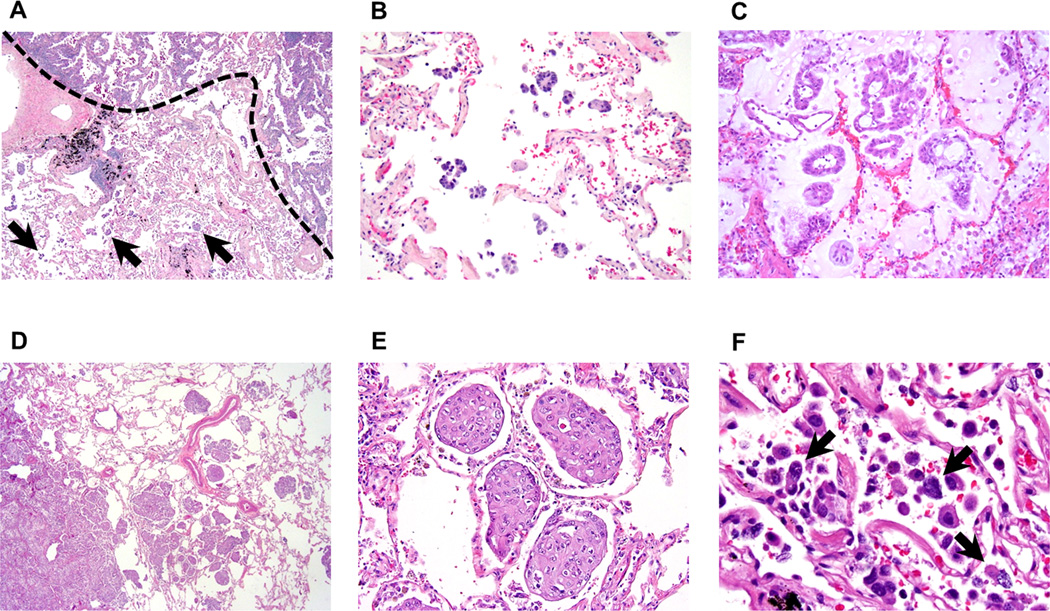
Figure 1. Morphologic features of tumor spread through air spaces (STAS) pattern (original magnification: ×20 in A and D; ×200 in B, C and E; ×400 in F).
(A) Micropapillary pattern STAS (arrows) identified within air spaces in the lung parenchyma beyond the edge (a dotted line) of the main tumor. (B) Micropapillary pattern STAS consisting of papillary structures without central fibrovascular cores. (C) Micropapillary pattern STAS forming ring-like structures within air spaces. (D) Solid pattern STAS identified within air spaces in the lung parenchyma beyond the edge of the main tumor. (E) Solid type STAS consisting of solid collections of tumor cells filling air spaces. (F) Single cell pattern STAS consisting of scattered discohesive single cells (arrows).

