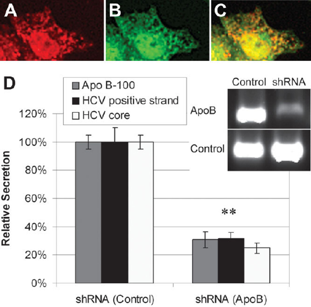
Fig. 2.
Double immunofluorescence staining of JFH-1–infected Huh7.5.1 cells. (A) Staining for HCV core protein (red). (B) Staining for ApoB100 (green). (C) Superpositioning of the images demonstrates that HCV core protein associates with ApoB100 in the cytoplasm. (D) Relative secretion of ApoB, HCV-positive strand RNA, and HCV core protein in JFH-1–infected Huh7.5.1 cells following silencing of ApoB100 mRNA by SureSilencing shRNA transfection. **P < 0.01.

