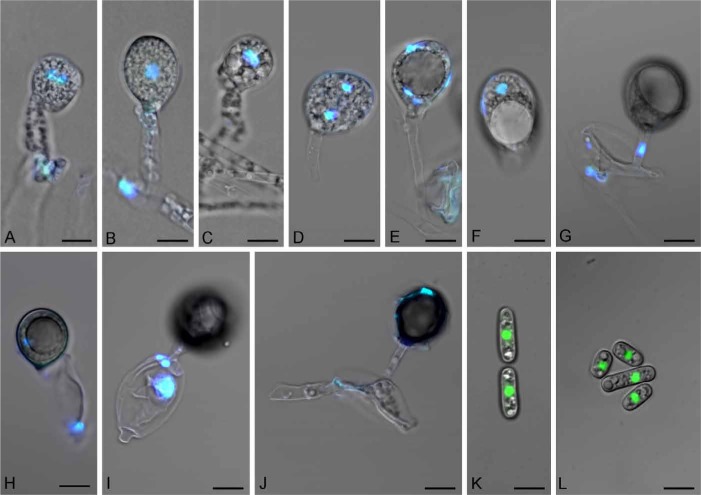
Fig. 1.
Basidioascus undulatus (DAOM 241956) sexual and asexual structures stained with DAPI (A–J) and SYTO 9 (K–L) and imaged with confocal microscopy. A. Dikaryotic basidium (2 nuclei). B. Karyogamy. C. Anaphase I. D. Telophase I and collapsed basal lateral projection. E. Telophase II (4 nuclei). F. Ejected basidium (4 nuclei). G–H. Maturation of a basidiospore on a basidium and the migration of a nucleus through the sterigma and into the basidiospore. I. Collapsing basidium with the three remaining nuclei. J. A totally collapsed basidium and probably one nucleus inside the mature basidiospore. K–L. Single nuclei inside arthroconidia. Bar = 5 μm.

