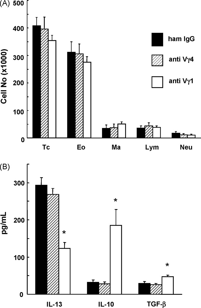
Fig. 1. Inflammatory cell infiltrate and cytokines in the airways of OVA-sensitized and challenged C57BL/6 mice treated with antibodies against TCR-γδ.
A. Mice were sensitized and challenged with OVA (2ip3N protocol). They were further treated twice with anti TCR-Vγ1 mAb, anti TCR-Vγ4 mAb or nonspecific hamster IgG (sham treatment) by i.v. injection 3 days before each of the two sensitizations. Inflammatory cells in BAL fluid were counted 48 h after the last OVA challenge. TC, total cells; Eo, eosinophils; Ma, macrophages; Lym, lymphocytes; Neu, neutrophils. Results for each group are expressed as the mean ± SEM (n = 12 in each group).
B. Mice were treated and BAL fluid collected as described under A. Cytokine assays were performed as indicated, and results for each group expressed as the mean ± SEM (n = 12 in each group). Significant differences (p < 0.05) between the anti TCR Vγ1 mAb-treated group and the other groups are indicated by *.

