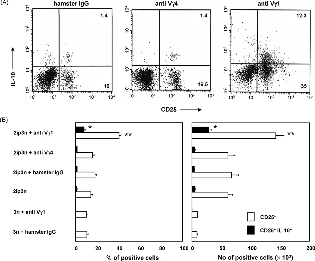
Fig. 2. Pulmonary CD4+CD25+ cells of OVA-sensitized and challenged C57BL/6 mice treated with antibodies against TCR-γδ.
A. Mice were treated as described in Fig.1. Pulmonary cells recovered 48 h after the last OVA challenge were stimulated with PMA/ionomycin in the presence of brefeldin A, and then stained with antibodies specific for CD4, CD25 and intracellular IL-10, as described in Materials and Methods. For three-color fluorescent analysis, cells were gated based on their scatter profile (lymphocyte gate) and on CD4-expression. 2.5 × 10⁴ gated cells were counted in each histogram.
B. Cell frequencies were determined cytofluorimetrically as described in 2A. Absolute cell numbers were obtained by multiplying the cell frequencies (percentage of CD4+CD25+ or CD4+CD25+ IL-10+ cells) by the number of cells obtained in lung digests. Results for each group are expressed as mean ± SEM (n = 12 in each group).

