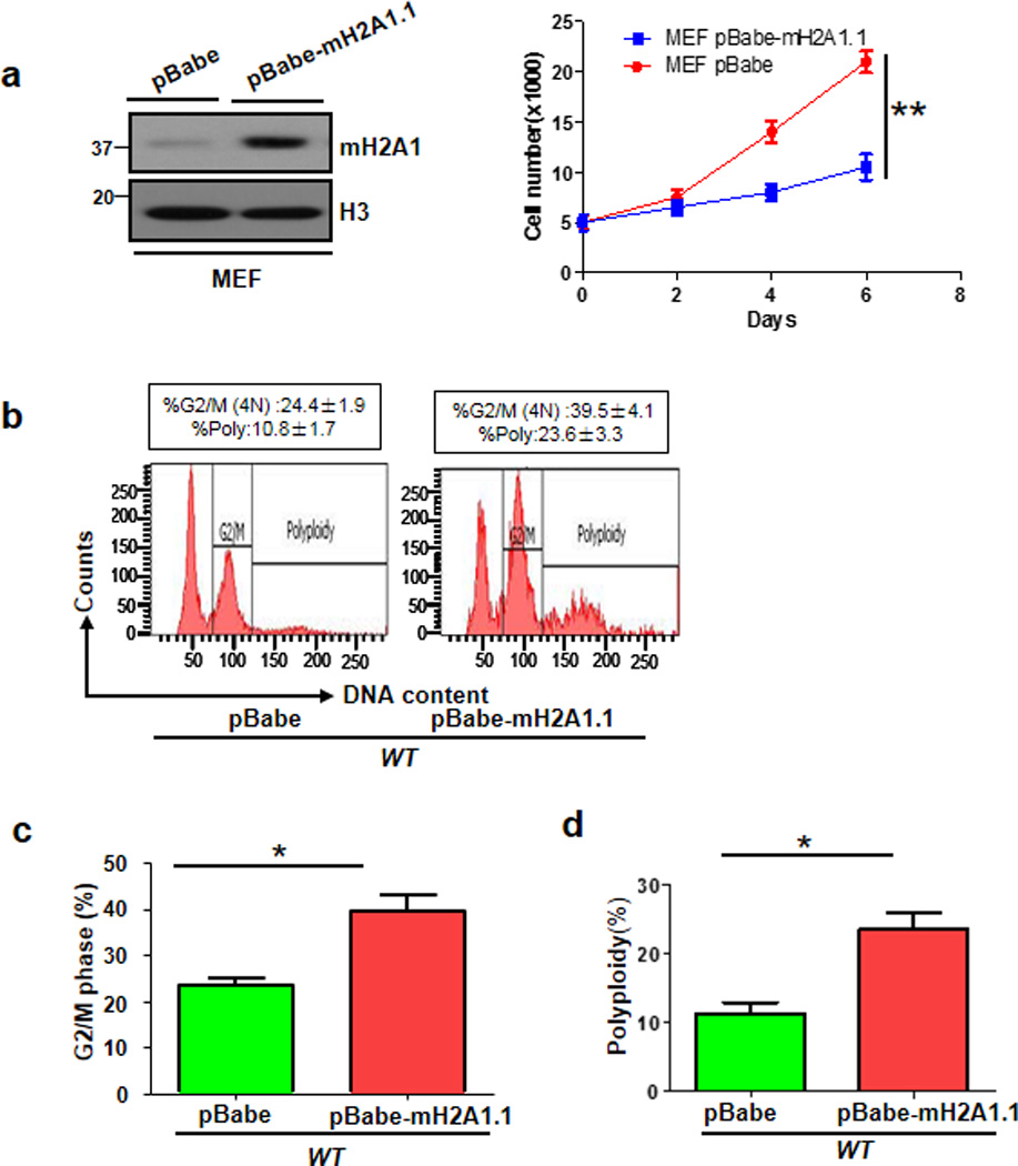
Figure 3. mH2A1.1 overexpression in primary MEFs triggers cell growth arrest and polyploidy.

(a) Primary MEFs were infected with pBabe or pBabe-mH2A1.1 lentiviral RNAs, selected, and harvested for Immunoblotting (Left panel). These cells were plated in 24-well plated for cell growth assay using direct cell counting (Right panel). (b) Flow cytometry analysis of DNA content in primary MEFs stably expressing pBabe or pBabe-mH2A1.1. (c) G2/M phase was determined by Flow cytometry analysis of primary WT MEFs with stably expressing pBabe or pBabe-mH2A1.1. The quantified results are presented as means ± s.d. (d) Polyploidy of primary WT and Skp2−/− MEFs with stably expressing pBabe or pBabe-mH2A1.1. The quantified results are presented as means ± s.d. (Error bars indicate s.e.m. Data represent mean values of three independent experiments. Student’s t-test used; *p<0.05;**p<0.01)
