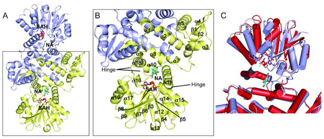
Figure 2.

Cartoon representations of NcsB1. (A) Dimer of NcsB1 with the active site region indicated. Chain A is colored light blue and chain B yellow. Ligands are depicted in stick format with SAH in red and naphthoic acid 4 in cyan. B) Close up view of monomer with secondary structural elements and hinge regions labeled. C) Overlay of NcsB1/SAH/4 monomer (light blue) and NcsB1/SAH monomer (red).
