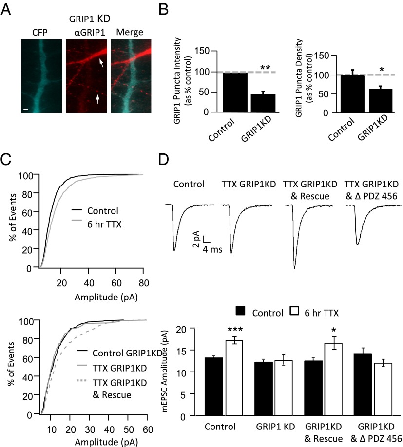
Fig. 4.
GRIP1 KD blocks synaptic scaling. (A) Sparse transfection of cultured neurons with the GRIP1 shRNA and soluble CFP (blue) (to label transfected neurons and dendrites) and αGRIP1 (red); the dendrite from the hairpin-expressing neuron shows little GRIP1 signal relative to the nontransfected dendrite (arrows). (B) Puncta Intensity (Left) and length density (puncta per μm) (Right) of GRIP1 puncta for control or shRNA-transfected dendrites; values from GRIP1KD neurons expressed as percentage of control. (C) Cumulative histograms of mEPSC amplitude from neurons in control and TTX conditions (Upper) and control GRIP1KD, TTX GRIP1KD, and TTX GRIP1KD & Rescue (Lower). (D) Average mEPSC waveform (Upper) and average mEPSC amplitudes from indicated conditions (Lower). (Scale bar: 1 µm.) *Different from control: P < 0.05; **P < 0.001.

