Fig. 2.
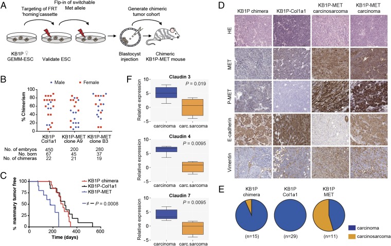
MET expression in KB1P chimeric mice leads to accelerated mammary tumor development. (A) Overview of the experimental procedure. Female ESCs derived from our KB1P mouse model were equipped with a Col1a1-frt homing cassette, and subsequently the Met proto-oncogene was introduced by recombinase-mediated cassette exchange. (B) Scatter plot illustrating the performance of KB1P ESC clones targeted with the Col1a1-frt homing cassette (Col1a1), and additionally equipped with Met (MET clone A9 and B3) to produce chimeric mice. (C) Mammary tumor-free survival of KB1P chimera (red; t50 = 284 d, n = 13 mice), KB1P-Col1a1 (black; t50 = 299 d, n = 14 mice), and KB1P-MET (blue; t50 = 176 d, n = 11 mice) chimeric mice. Log-rank (Mantel–Cox) P values are indicated. (D) Immunohistochemical staining of formalin-fixed/paraffin-embedded sections from KB1P, KB1P-Col1a1, and KB1P-MET chimeric mammary tumors for H&E, MET, METY1234/1235, E-cadherin, and vimentin. (E) Pie diagrams illustrating the distribution of different mammary tumor types in KB1P (93% carcinoma; 7% carcinosarcoma), KB1P-Col1a1 (100% carcinoma), and KB1P-MET (45% carcinoma; 55% carcinosarcoma) chimeric mice. Carcinomas are shown in blue and carcinosarcomas in orange. (F) Box plot showing relative expression of claudin 3, 4, and 7 in KB1P-MET carcinomas and carcinosarcomas. Mann–Whitney P values are indicated.

