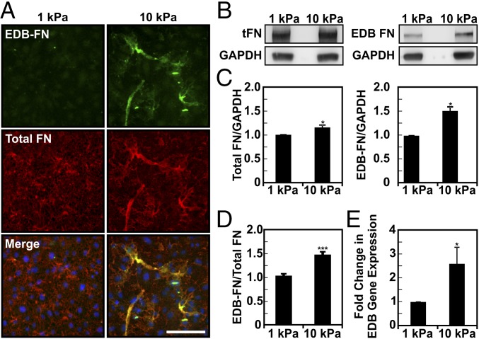
Fig. 3.
Expression of the EDB-FN splice variant increases with ECM stiffness in vitro. ECs were plated on compliant (E = 1 kPa) and stiff (E = 10 kPa) substrates (100,000 cells/mL), grown over 3 d, and subjected to immunofluorescence staining or Western blot. (A) Immunofluorescent staining of both EDB-FN and total FN increases with increasing substrate stiffness (images were acquired with the same exposure settings). (Scale bar, 50 μm.) (B) Representative Western blot of total protein extracts showing EDB-FN and total FN on compliant and stiff substrates. GAPDH was used as loading control. (C) Densitometric quantification of EDB-FN and total FN normalized against GAPDH content. (D) Corresponding ratio of EDB-FN to total FN showing a 30% increase in EDB-FN expression with increasing stiffness (three independent experiments). (E) EDB-FN expression as determined by quantitative real-time RT-PCR indicates a significant increase in mRNA expression with increasing stiffness (three independent experiments). Plots are mean ± SE, Student t test: *P < 0.05, ***P < 0.001.

