Fig. 4.
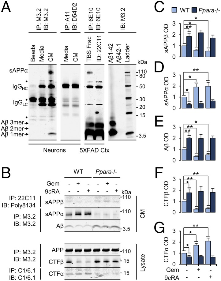
Activation of PPARα attenuates endogenous Aβ generation. (A) Representative immunoblots of APP species IP from 18DIV neuronal culture media using antibodies M3.2 and A11 or from the hippocampal TBS fraction (TBS Frac) of 6-mo-old 5XFAD mice with antibody 6E10. (B–G) Representative immunoblots (B) and quantification of murine sAPPβ (C), sAPPα (D), and Aβ (E) immunoprecipitated from conditioned media, and β-secretase cleaved APP C-terminal fragments (CTFβ) (F) and α-secretase cleaved APP C-terminal fragments (CTFα) (G) immunoprecipitated from lysates harvested from WT and Ppara−/− neurons treated with DMSO, 25 µM GEM, or 0.2 µM all-trans retinoic acid (9cRA) for 24 h. All values are corrected for IgG, indicate the mean ± SEM relative to control, and represent n = 3 or 4 for each treatment. *P < 0.05 and **P < 0.01, using two-way ANOVA. IB, immunoblot; Beads, protein G beads alone; IgGHC/LC, IgG heavy chain and light chain; Media, unconditioned media; OD, relative optical density. ●, nonspecific band.

