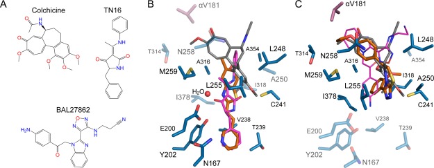
Figure 3.

Comparison of MI-181 and C2 binding with colchicine, TN16, and BAL27862 binding on β-tubulin. A: The chemical structures of colchicine, TN16, and BAL27862. B: The MI-181 (orange) binding site with tubulin-bound colchicine (gray) and TN16 (magenta) superimposed using the β-subunit. Residues interacting with MI-181 are shown as sticks and labeled. Residues that interact with C2 are highlighted with transparency. C: As in B, the C2 (orange) binding site superimposed with colchicine (gray) and BAL27862 (magenta). Residues that interact with MI-181 are transparently depicted.
