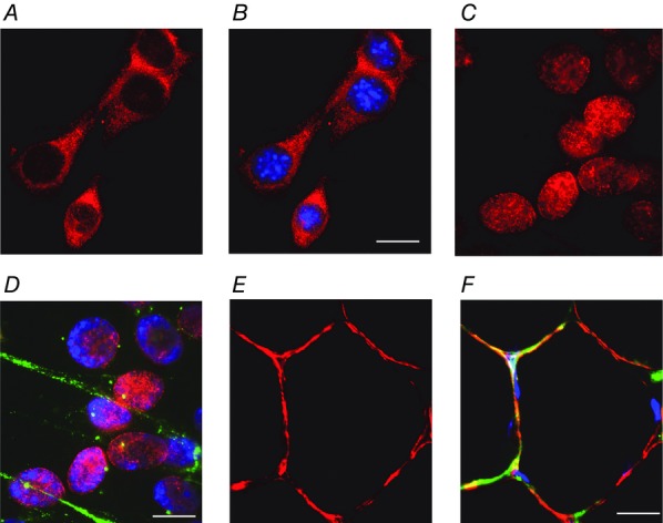
Figure 1.

The localisation of GRs changes with muscle differentiation
Photographs showing the expression and localisation of GRs (red), laminin (green) and double stranded DNA (nucleus; blue) in myoblasts (A and B), myotubes (C and D) and muscle fibres (E and F). Note that the GRs are expressed mainly in the cytoplasm in myoblasts, in the nucleus in myotubes, and in a narrow band along the cell surface in adult muscle fibres. Scale bars: A and B, 5 μm; C and D, 2.5 μm; E and F, 15 μm.
