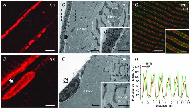
Figure 2.

GRs are localised in the extracellular matrix and around satellite cells in adult mouse muscle fibres
A and B, immunofluorescence confocal microscopy images showing the localisation of GRs in mouse soleus. Note that the GRs are localised either in the proximity of the surface membrane, where they appear to form a narrow and discontinuous band (A; white dashed square) and in satellite cells (B, white arrow) or in the fibre interior where the signal is mostly localised in the I-band. C, D, E and F, immunogold-labelling electron microscopy photographs showing the localisation of GRs in mouse slow-twitch fibres. Note that the GRs (dark dots, i.e. gold particles) are abundant in the extracellular matrix (C and enlargement of the dotted box in D) outside the sarcolemma (C, filled arrow) and in satellite cells (E, open arrow). GRs are also present within the fibre interior almost exclusively at the I-band, and mostly in the proximity of mitochondria (C, E and F). G, a confocal micrograph showing a muscle section immunoblotted for both GRs and RYR1s. Note that the GRs (red) are mostly spread throughout the I-band inside the double green lines generated by the staining of RYR1s, which marks the position of triads. H, graph representing the fluorescence intensity profile calculated from images obtained from samples co-immunostained for RYR1s and GRs. Scale bars: A, 5 μm; B, 10 μm; C and E, 5 μm; D and F, 0.5 μm; G and inset, 10 μm and 1 μm, respectively.
