
Figure 1.
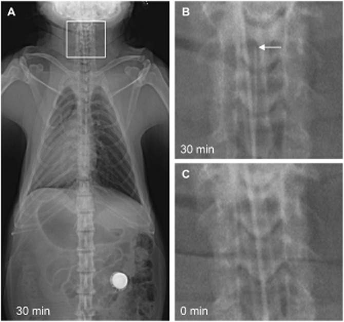
Representative radiographic images of a catheterized monkey with the catheter tip located in the cisterna magna (C1-2). (A) Image in ventral-dorsal projection taken 30 min after administration of Omnipaque 300 contrast. (B) Zoomed-in image of the boxed area in panel A showing the catheter tip with an arrow. (C) Image taken immediately before administration of the contrast as a baseline control.