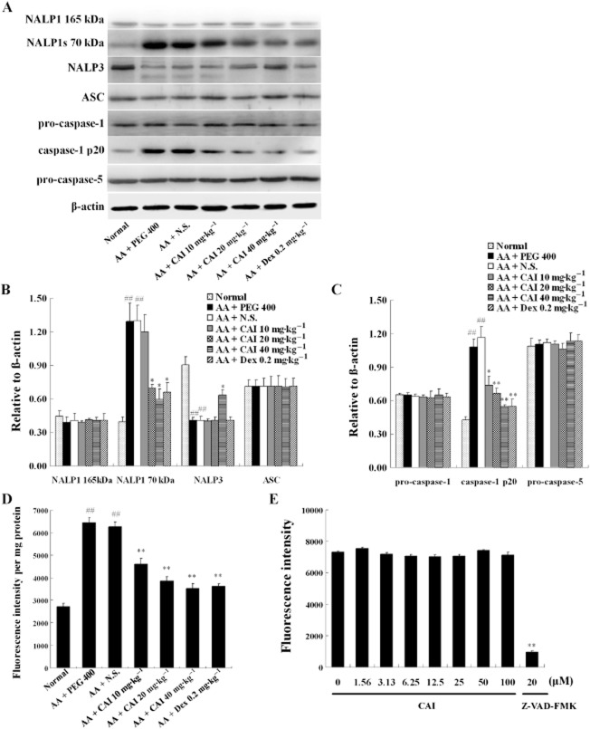
Figure 6.
Detection of the components of inflammasomes in the synovium and the effects of CAI. (A) Synovial tissues were homogenized and analysed by Western blot, with the antibodies as indicated. (B and C) Densitometric analysis of immunoblot bands. β-actin was used as the internal control. Data shown are means ± SEM of three rats. (D) Caspase-1 activity was measured in synovial tissues from rats from different groups. Data shown are means ± SEM of four rats. ##P < 0.01 versus normal group, *P < 0.05, **P < 0.01 versus vehicle-treated AA group. (E) The direct effects of CAI on the activity of caspase-1 in vitro were assayed. The specific caspase-1 inhibitor Z-VAD-FMK was used as positive controls. Data shown are means ± SEM from six independent experiments. **P < 0.01 versus the control without drug (0 μM).

