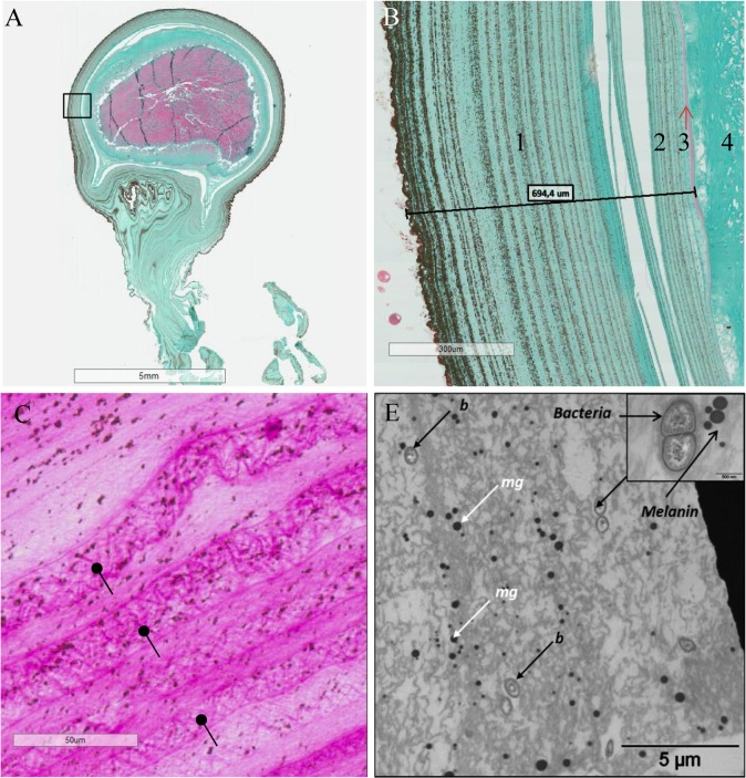
Fig 1. Egg Case and SepECP structure analysis by histological techniques.
(A) Longitudinal section of a whole 15-day-old egg. Black box, magnificence zone of Fig 1.B. (B) Longitudinal section of a 15-day-old egg stained in Prenant-Gabe trichromic. 1: outer layer of the extrachorion, 2: inner layer of the extrachorion, 3: chorion, 4: vitelline membrane, 5: oocyte. (C) Longitudinal section of a 24-hour-old egg focused on the outer layer of the extrachorion stained in Periodic Acid of Schiff. Glycoproteins polymerize and are assembled in narrow meshes (black arrows) that are organized in layers. (D) Section of the outer layer of the extrachorion of an egg in TEM (b, bacteria; mg, melanin granules).

