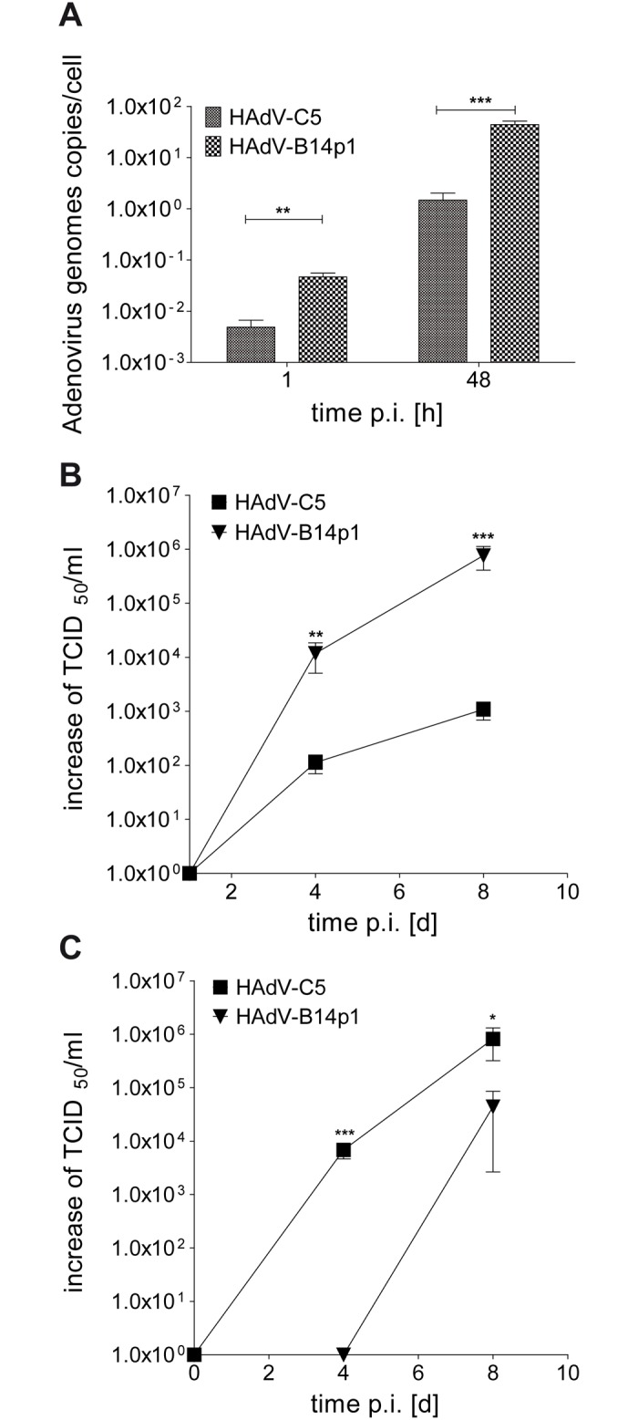
Fig 3. Efficient apical infection of differentiated human bronchial epithelial cells with HAdV-B14p1.

(A) Intracellular HAdV genomes were quantified by qPCR at 1 h and 48 h p.i. (** p< 0.01; *** p< 0.001, unpaired t-test) (B) Release of infectious virus progeny at the apical side of the differentiated bronchial epithelial cell layer as determined by the TCID50 method on day 1, 4, and 8 p.i. (** p< 0.01; *** p< 0.001, unpaired t-test). (C) Release of infectious virus progeny at the basal side of the differentiated bronchial epithelial cell layer as determined by the TCID50 method on day 1, 4, and 8 p.i. (* p< 0.05; *** p< 0.001, unpaired t-test). The TCID50 values in B and C are normalized against the input virus titers measured on day 1 p.i. and set to 1 x 100 because the TCID50 values measured on day 1 are probably remaining viral particles originating from the virus inoculum.
