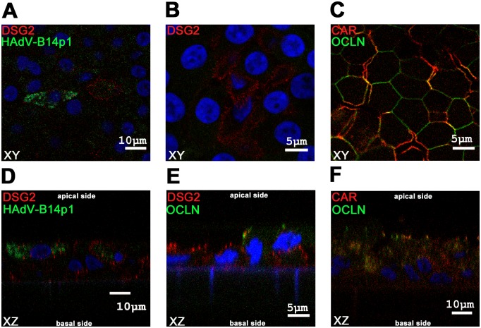
Fig 4. Immunofluorescence staining of differentiated human bronchial epithelial cells.
Confocal microscopy immunofluorescence analysis of differentiated human bronchial epithelial cells. Figs A, B, C show XY planes, Figs D, E, F show XZ planes. (A, D) Cells were infected from the apical side with HAdV-B14p1 at a moi of 10 (TCID50/cell), fixated 4 days p.i. and stained for HAdV in green (FITC conjugated antibody) and the desmoglein 2 (DSG2 receptor) in red (dsRed antibody), the nucleus was counterstained in blue (DAPI). (B, E) Differentiated human bronchial epithelial cells stained for DSG2 receptor in red (dsRed) and occludin (OCLN) a tight junction marker in green (FITC), nucleus was counterstained in blue (DAPI). (C, F) Differentiated human bronchial epithelial cells were stained for CAR receptor in red (dsRed) and the tight junction marker OCLN in green (FITC) and the nucleus in blue (DAPI).

