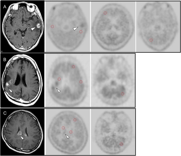
Fig 1. (A) 60-year-old male with recurrence of brain metastasis (lung carcinoma) at left temporal lobe (arrow head).
MET uptake of the MRI-enhanced lesion showed higher than the region around the lesion, the contralateral brain, the contralateral frontal lobe and cerebellum. (B) 26-year-old male with recurrence of an anaplastic astrocytoma in the right temporal lobe. Contrast-enhanced MRI showed gadolinium-enhanced nodular lesion (arrow) in the right temporal lobe, which was the location of post-surgical resection of the primary brain tumor. MET uptake of the MRI-enhanced lesion (arrow head) showed higher than the region around the lesion, the contralateral brain, the contralateral frontal lobe and cerebellum. (C) 59-year-old female with radiation necrosis. Contrast-enhanced MRI showed gadolinium-enhanced lesion (arrow head) at the corpse callosum, which was suspected recurrent brain metastasis (lung carcinoma). MET uptake of the MRI-enhanced lesion showed slightly higher than the region around the lesion, the contralateral brain, and the contralateral frontal lobe, but similar to MET uptake at cerebellum.

