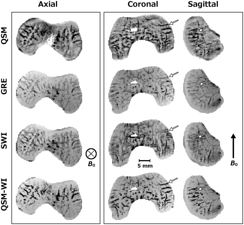
Fig 4. Comparison of QSM, plain GRE, SWI and QSM-WI at 7.0 T ex vivo.
Comparison of QSM post-processed, plain GRE, SWI and QSM-WI datasets from a 1-month-old human specimen scanned at 7.0 T (TE = 29.06 ms and bandwidth = 60 Hz/pixel) in 3 mm-thick mIPs with QSM contrast inverted to match SWI and GRE. The first pane shows the axial plane, perpendicular to B 0. All four techniques demonstrated closely similar results. In the planes parallel to B 0 (second pane), GRE and QSM demonstrated a closely similar visual appearance; however, the splitting artifact along B 0 was evident in the SWI post-processed data. QSM-WI demonstrated both corrected artifacts and improved visualization of the cartilage canals.

